Heart & Aorta in PA View: Anatomy & Imaging Essentials
Generated from prompt:
posterior anterior position of heart and aorta describe position ,direction and image essential in detail ( slide no should be 1-5 if any anatomy is related to it then also add it
This deck details the posterior-anterior positions, orientations, and key imaging features of the heart and aorta in PA chest X-rays, including borders, silhouettes, relations to lungs, spine, esophagus, and normal anatomy for radiology and anatomy.
Slide 2 - Heart Position in PA View
- Located in middle mediastinum: ~2/3 posterior to sternum, ~1/3 anterior to vertebral column
- Apex directed inferiorly, anteriorly & leftward (left 5th ICS, mid-clavicular line)
- Base (atria/posterior) faces posteriorly toward esophagus/spine
- Long axis obliquely tilted ~45° leftward/inferiorly
- Borders in PA: Right=SVC/RA; Left=LV/aortic arch; Superior=appendages; Inferior=RV
Source: Wikipedia: Cardiac anatomy
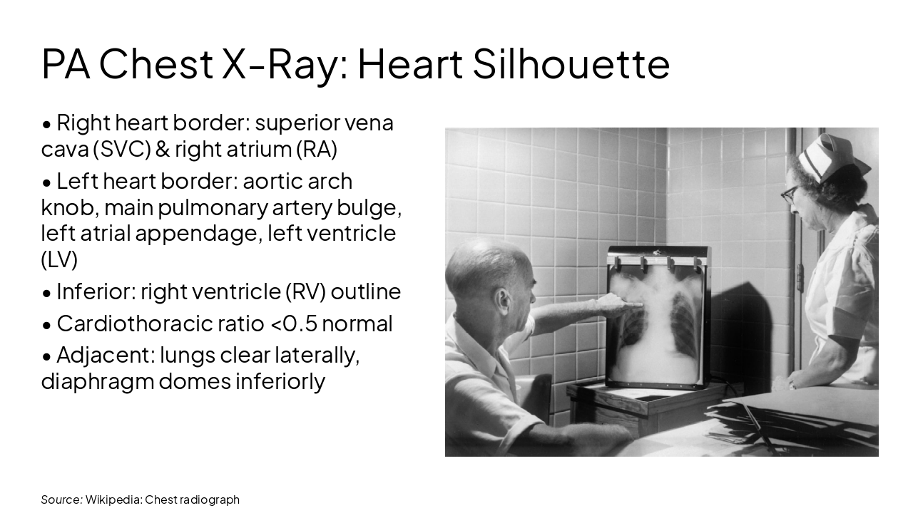
Slide 3 - PA Chest X-Ray: Heart Silhouette
- Right heart border: superior vena cava (SVC) & right atrium (RA)
- Left heart border: aortic arch knob, main pulmonary artery bulge, left atrial appendage, left ventricle (LV)
- Inferior: right ventricle (RV) outline
- Cardiothoracic ratio <0.5 normal
- Adjacent: lungs clear laterally, diaphragm domes inferiorly
---
Source: Wikipedia: Chest radiograph
Slide 4 - Aorta Position & Direction in PA View
- Ascending aorta: posterior to lower sternum, directed superiorly & slightly rightward
- Aortic arch: convex leftward at T4 level (aortic knob visible left superior mediastinum)
- Arch branches: brachiocephalic, left common carotid, left subclavian
- Descending thoracic aorta: posterolateral to esophagus, descends along left spine side to T12 hiatus
- Overall PA projection: arch prominent, ascending obscured by spine/heart, descending faint left paravertebral
Source: Wikipedia: Aorta anatomy
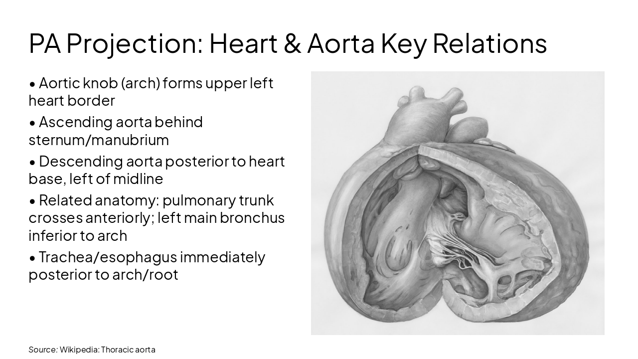
Slide 5 - PA Projection: Heart & Aorta Key Relations
- Aortic knob (arch) forms upper left heart border
- Ascending aorta behind sternum/manubrium
- Descending aorta posterior to heart base, left of midline
- Related anatomy: pulmonary trunk crosses anteriorly; left main bronchus inferior to arch
- Trachea/esophagus immediately posterior to arch/root
---
Source: Wikipedia: Thoracic aorta
Discover More Presentations
Explore thousands of AI-generated presentations for inspiration
Create Your Own Presentation
Generate professional presentations in seconds with Karaf's AI. Customize this presentation or start from scratch.