Slide 1 of 6
Slide 1 - Clinical Case Review
Clinical Case Review
Case Presentation: Autoimmune Pancreatitis vs. Pancreatic Adenocarcinoma

Generated from prompt:
Redesign the uploaded PowerPoint into a modern, sleek, classy, and smooth clinical case presentation. STRICT RULES: - Do NOT modify the first slide at all. - Do NOT change ANY text, wording, punctuation, or content on any slide. - Only improve visual design, layout, typography, spacing, hierarchy, and color usage. DESIGN STYLE: - Minimalist medical theme (clean, professional, elegant) - Use a neutral palette (white, deep blue, subtle gray accents) - Strong typography hierarchy (titles bold, body readable, key data emphasized) - Increase whitespace and readability - Convert dense paragraphs into visually structured layouts (but WITHOUT changing text) - Align content cleanly (grid-based layout) - Use subtle section dividers and consistent slide structure - Improve tables and comparisons visually (clear contrast, alignment, spacing) - Highlight key clinical data (IgG4, CA19-9, imaging findings) using visual emphasis (color or weight, not extra text) - Keep animations minimal or none SPECIFIC IMPROVEMENTS: - Case presentation slides: better spacing, bullet alignment, and emphasis on key findings - Biology and imaging slides: structured layout for readability - Comparison tables (PAI vs ADK): modern table styling, clear contrast - Diagnostic reasoning slides: emphasize decision flow visually - Avoid clutter and over-design OUTPUT: - Modernized PPTX presentation - Same slide order and exact content preserved - Clean, consistent slide master applied throughout Goal: Make it look like a high-end conference presentation without altering scientific content.
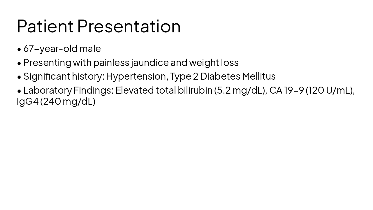
This deck presents a clinical case of a 67-year-old male with painless jaundice and pancreatic mass, differentiating autoimmune pancreatitis (PAI) from pancreatic adenocarcinoma (ADK) through patient history, labs (IgG4, CA 19-9), imaging, diagnostic
Clinical Case Review
Case Presentation: Autoimmune Pancreatitis vs. Pancreatic Adenocarcinoma
| Feature | Autoimmune Pancreatitis (PAI) | Pancreatic Adenocarcinoma (ADK) |
|---|---|---|
| Onset of Symptoms | Subacute, often vague | Rapid, progressive |
| CA 19-9 Levels | Normal or mildly elevated | Significantly elevated |
| IgG4 Levels | High (>135 mg/dL) | Normal |
| Imaging Findings | Diffuse enlargement, capsule sign | Focal mass, ductal cutoff |
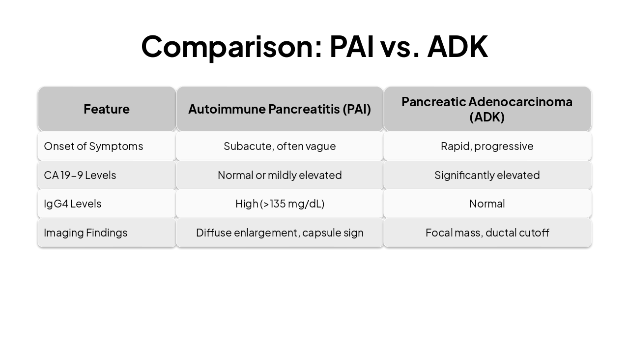
Autoimmune Pancreatitis remains a crucial differential diagnosis in pancreatic mass presentations requiring integrated diagnostic approaches.
Clinical Case Presentation Summary
Explore thousands of AI-generated presentations for inspiration
Generate professional presentations in seconds with Karaf's AI. Customize this presentation or start from scratch.